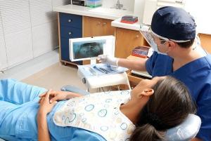
During the past decade, prosthetic dentistry has positively been shaped and impacted by computer-driven technologies that situate a level of desirable comfort and efficacy for the dentist and patients alike. Among many biometric systems implemented in the two cardinal fields of prosthodontics and implantology, rehabilitation of partially or fully edentulous patients has become substantially easier and precise. Fabrication of dentures and other prostheses by means of computer-aided design (CAD) and computer-aided manufacture (CAM) has not only proven to be possible but may have readily revolutionized the world of dental prosthetics. With the rapid integration of technology for prosthetic marketing, dentists have been adopting a high-yielding system of immediate dental restorations based on digital impressions and workflow that prioritizes patient control and convenience.
What is a dental impression?
A conventional dental impression is a negative imprint of the hard and soft tissues in the mouth, recording of which can generate a positive reproduction (cast or model). It is a preliminary step in which a stock or custom dental impression tray is loaded with appropriate material (commonly used hydrocolloids like agar or alginate) that is specially designed to roughly engage the oral structures and fit over the dental arches. This impression is then aptly coerced into the fabrication of necessary dental appliances that fulfill a patient’s dental needs.
The principle of impression-taking abides by either of these two theories of denture appliance fabrication- the mucostatic theory and the mucocompressive theory. Mucostatic theory follows the impression being taken of the mucosa when in its normal resting position. Although these impressions are generally affiliated with great fit and accuracy, the denture is more likely to pivot around compressive areas such as the torus palatinus, during chewing. Mucocompressive theory, on the other hand, is related to taking impressions when the mucosa is subject to compression. As a result, a denture most stable at function but not at rest is accomplished. Another type of impression technique, known as the select pressure technique is focused on delivering relief to stress relief areas and compression to the stress-bearing areas. This guarantees the benefits of both the theories; the best of both worlds.
What are digital impressions?
A digital impression is a virtual scan that creates a map of the oral cavity. Digital impressions optimize cutting-edge technology that allows dentists to create a computer-generated virtual replica of the hard and soft tissues of the oral cavity using lasers and other optical scanning devices. The digital technology captures a coherent and structured impression of the oral tissues that can then be transmitted to computer software. This analyzes the data and lets the dentist implement modifications to create dental appliances that have superior fit and precision. A milling unit or fabrication plant will then fabricate a physical model of the crowns, bridges, dentures, or other restorations that would be given to the patient.
A majority of patients seeking prosthodontic rehabilitation have edentulous ridges, whether partial or complete. Digital impressions ensure a better fit for the underlying (potentially resorbed) tissues and the CAD/CAM system substitutes many of the steps of a conventional denture production chain. For instance, occlusal rims (OR) and functional impression with border moldings can be digitized, with the help of intraoral scanners, thus reducing the number of appointments for the patient till a final denture delivery.
Types of digital impressions technology
The innovative technology incorporated in digital impression-taking can be availed of by the dentist in two major ways. One type captures the images as digital photographs, and the other captures images as digital video. Since the images can be readily produced in a matter of seconds, it allows dentists and laboratory technicians to view and modify a series of images to create a denture fit that is accurate to the particular patients.
The images can be captured either using lasers or digital scanning. Laser scanners use concentrated light to capture all the details of the teeth and gums. They are safe and highly precise and will help eliminate the patient’s need to hold distasteful material in his or her mouth as would with the conventional method of impression-taking. An intraoral scanner is a handy device that works by projecting a light source onto the object to be scanned (in this case, the alveolar ridges or prepared teeth). They may, however, require powder-coating before scanning to ensure complete inclusion of the oral structures, although some modern-day scanners work without powder use.
How is a digital impression created?
Contrary to popular belief, the digitization of impression-taking is not relevant in all cases, and most commonly may only be “partially digital”. Prior to a digital outcome, a preliminary impression through conventional methods is crucial. This is because although digital scanners are great at recording precise readings of hard tissues, they are not accurate in recording the compressed and relaxed states of soft tissues. Some clinical cases have shown the pathway until the delivery of prosthetic appliances utilizing only digital intraoral scans in a fully digital workflow (mostly without the introduction of border molding).
Studies have demonstrated that in such techniques, the denture is able to provide sufficient retention, however, the plica intermedia is usually overextended and the denture is not secured with the functional movements of the mouth, giving substandard results. Since they marginalize the functional mucosa movements, the reliability and reproducibility of some of these techniques are questionable. For this reason, end-to-end digital fabrications are usually foreshadowed by conventional impressions.
A denture prototype can be fabricated by additive manufacturing (AM) for the chairside try-in sessions. A final denture is manufactured using subtractive or additive manufacturing methods.
- The dentist prepares the patient’s tissues for scan by suctioning out any blood and saliva that may coat the teeth and other soft tissues with specially formulated titanium dioxide powder. Some scanners are powder-free.
- Using the intraoral scanner wand, the dentist captures a series of 3D digital images or videos that can be viewed on the monitor by the patient and the dentist.
- The affected area is shown attention by gliding the scanner to the site. The dentist inspects the area for resorbed tissues, uneven dental ridges, any early signs of oral trauma, oral lesions, and other anomalies that can deter the process of impression-taking.
- The buccal-occlusal-palatinal (BOP) and “zig-zag” techniques are mainly used to digitally scan the edentulous jaws. Using special markers on the jaws, the dentist is able to avoid “overlapping effects” of the scanned image or video.
- On the chairside screen, the dentist is able to view the impression data in image or video format that has been compiled by the software.
- The dentist then reviews the digital images and verifies the accuracy of the scan.
- The digital impressions are sent to the dental laboratory where the patient’s restorations are fabricated, finished, polished, and sent back to the patient. Some clinics may also choose to 3D print the dentures with the relevant data from the digital scans.
Benefits of digital impressions
The process of digital impression-taking and denture fabrication is ridden with amazing benefits:
- Supreme patient convenience: The process of impression-taking becomes powderless and seamless with the digital intraoral scanning technique.
- Impressive speed: No longer do you have to wait around for the impression to dry. With incredible scanning algorithm, the Medit i500 ensures time-efficiency.
- Enhanced accuracy: They can scan oral tissues to a T, irrespective of the most minute of oral structures or any oddities. They are brilliant equipment for taking measurements.
- Simplified dental procedures: In cases of multiple dental implants or complex undercuts, dental procedures are simplified with the Medit i500.
- Superior predictability: The implementation of facial scanning into the workflow along with intraoral scanning allows the dentist to provide a virtual try-in session. The patient is able to see how the outcome is mostly to look like.
- Improved patient communication: Patients are more satisfied with intraoral scanning as they feel more involved in their procedures
Patients receiving digitally fabricated dentures may be able to transition from a traditional denture to an immediate denture quicker and easier, with the likeness of brilliant precision, accuracy, and patient comfort. Furthermore, the facial scans using this digital prototype can be obtained for the virtual esthetic evaluation (by the dentist and the patient) and digital try-in session. Many patients find digital impressions to be more comfortable than traditional impression-taking, which consequently helps the dentist build a strong rapport and grow their practice extensively.